|
Ultrasound is a non-invasive procedure that uses soundwaves to provide moving pictures of your pet's organs. Through the use of ultrasound, we can:
- Obtain urinalyses via cystocentesis (directly from the bladder);
- Confirm the presence of bladder stones or abdominal fluid;
- Investigate the extent of a patient's kidney disease;
- Examine a patient's internal organs for irregularities;
- Verify abnormalities that we have viewed on a patient's abdominal radiographs. Ultrasound provides greater clarity to its images so that we can identify an abdominal mass, its location, and whether it is attached to any organs.
With the ability to obtain real-time information via ultrasound, the results can be evaluated immediately; however, in some cases the images might be sent to a veterinary radiologist for a more in-depth evaluation. Ultimately, the use of ultrasound will provide us greater opportunity to swiftly and accurately diagnose your pet in order to begin treatment as soon as possible.
This ultrasound image shows a tumor in the kidney of a 10-year-old cat. Our doctors were able to compare this to an abdominal radiograph in order to make an accurate diagnosis. Please click here to view this cat's radiograph.
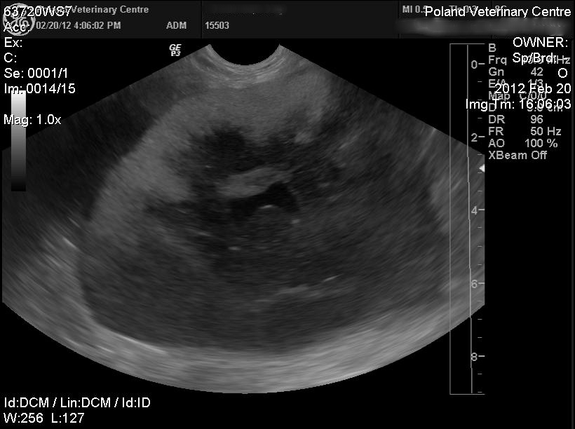
The following ultrasound image depicts sediment in a 5-year-old male cat's bladder. This type of sediment can lead to urinary obstruction, which can be very painful and require medical treatment.
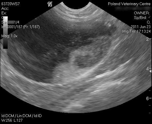
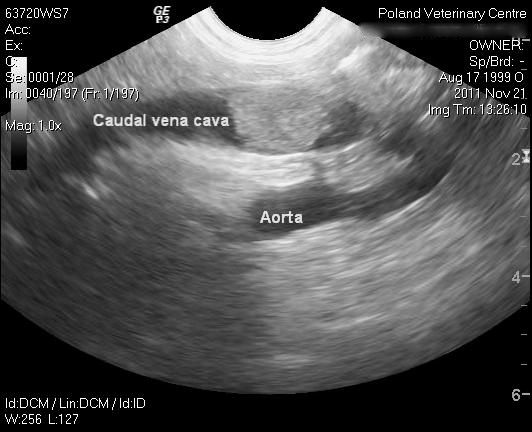
This ultrasound illustrates an adrenal tumor that has invaded the caudal vena cava of a 12-year-old male dog. Upon discovering this tumor, the doctors were able to discuss treatment options with the dog's owner.
|


