We would love to hear from you!
|
|
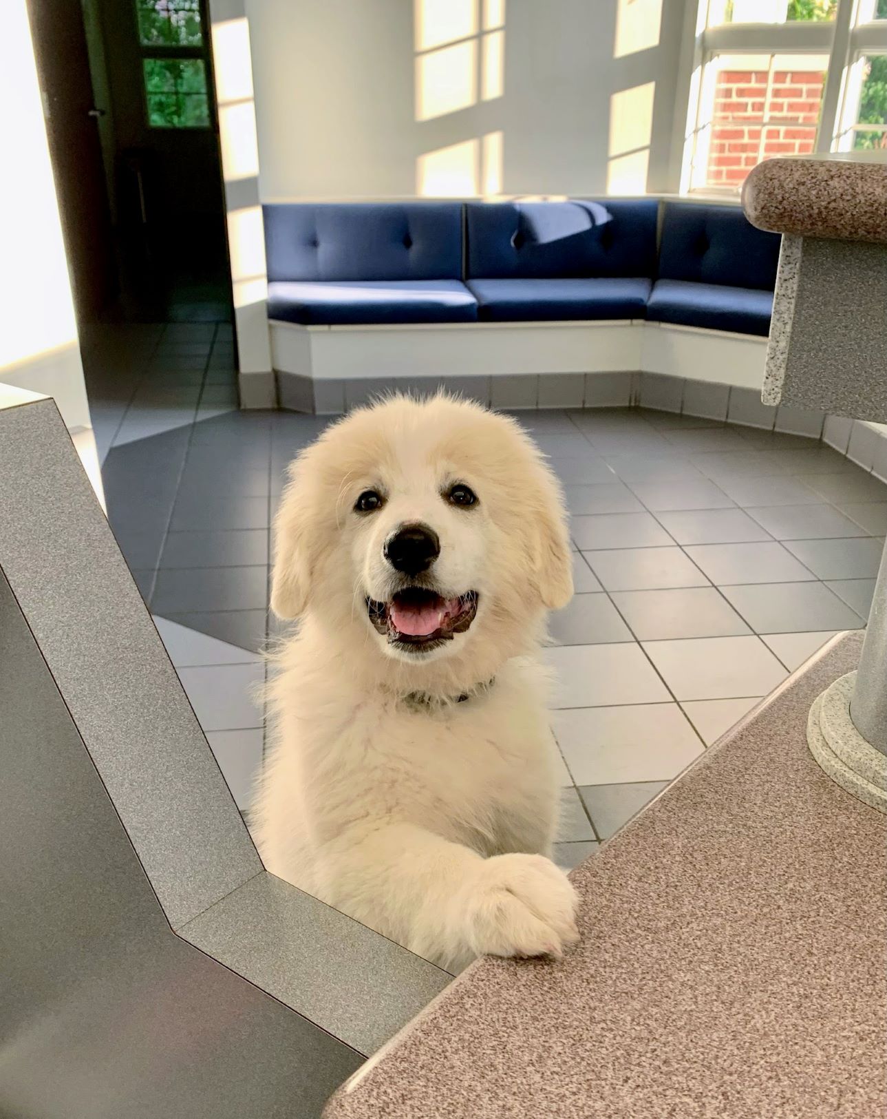
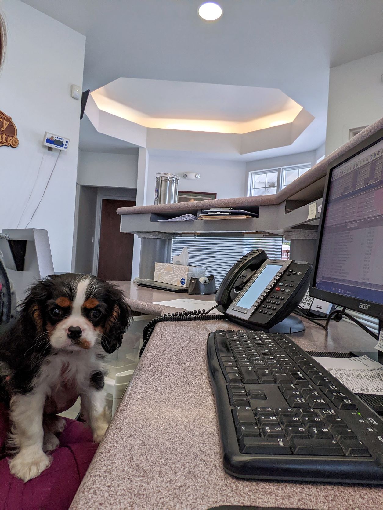
Welcome to Poland Veterinary Centre!
At Poland Veterinary Centre, we recognize the special relationship between people and their pets. We are a full-service small animal hospital in Poland, Ohio.
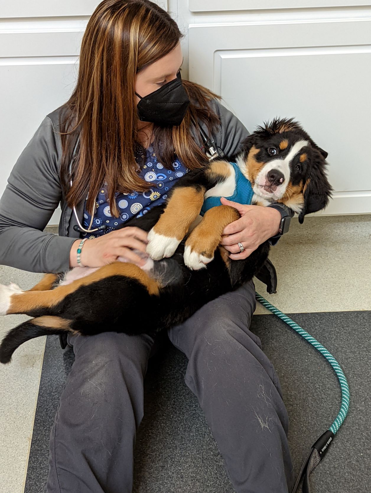
Our job is to work as a team to provide the best possible care to our patients. We educate our clients about caring for their pets and what veterinary medicine can do for them. We build relationships with our clients, and we show them how much we care through our interactions with their pets and their families. We are committed to being the best at what we do, and we strive to exceed our clients' expectations every day.
We want you and your pet to have a great experience at our hospital. Please see our Visit Preparation page by clicking here for helpful tips on getting the most out of your veterinary visit.
|


.jpg)


